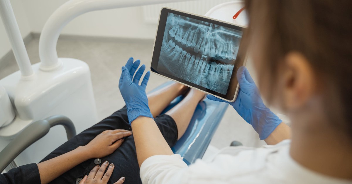
Why Dental X-Rays Should Always Be Done: Exploring the Benefits
Going to the dentist can be a stressful experience for many people, especially if they are unsure about the procedures involved. One such procedure is the dental x-ray, which is often performed during a routine dental examination. Some people may wonder if it is really necessary to have an x-ray taken, especially if they are concerned about radiation exposure. Let’s explore why dental x-rays should always be done during an examination and how to start your career in dental radiology.
How Dental X-Rays Work
Dental x-rays use electromagnetic radiation to produce images of the teeth and surrounding tissues. The x-ray machine emits a beam of radiation that passes through the mouth and is absorbed by the bones and tissues in its path. The remaining radiation reaches a sensor or film, which captures the image. The resulting image shows the dentist the internal structure of the teeth, including any decay or abnormalities that may not be visible to the naked eye.
The Purpose of a Dental X-Ray
The purpose of a dental x-ray is to help the dentist diagnose any issues with the teeth and surrounding tissues. X-rays can detect cavities, bone loss, tumors, cysts, and other abnormalities that may be hidden from view. By identifying these problems early, the dentist can develop a treatment plan that will prevent further damage and preserve the patient’s oral health.
Are There Restrictions on Who Can Have an X-Ray?
While dental x-rays are generally safe for most patients, there are some restrictions on who can have them. Pregnant women, for example, should avoid x-rays unless absolutely necessary, as the radiation can harm the developing fetus. Patients with certain medical conditions, such as thyroid problems or cancer, may also need to avoid x-rays or take special precautions. Your dentist will review your medical history and determine if an x-ray is appropriate for you.
How Frequently to Have a Dental X-Ray
The frequency of dental x-rays depends on the individual patient’s needs. Patients with a high risk of dental problems, such as those with a history of cavities or gum disease, may need to have x-rays taken more frequently than those with a lower risk. In general, most patients should have x-rays taken every one to two years to monitor their oral health.
Pre and Post Dental X-Ray
Before a dental x-ray, the patient will be asked to remove any jewelry or metal objects from their mouth, as these can interfere with the image. The patient will then be positioned in front of the x-ray machine, and the dentist or radiology technician will take the image. After the x-ray, the patient can resume their normal activities immediately.
Enroll Today to Study Dental Radiology at The Westchester School for Medical and Dental Assistants
If you are interested in learning more about dental radiology, consider enrolling in a program at The Westchester School for Medical and Dental Assistants. Our comprehensive program will prepare you for a career as a dental radiology technician, where you can help dentists diagnose and treat a variety of oral health issues. Get in touch with us to learn more!